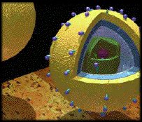
 An early understanding of the HIV virus
3D Computer-Assisted
Animated
Models and Animations
![]()
Produced in
Softimage, Eddie, and Amazon:
3D Animation,
Texture, Bump, Displacement and Distortion Maps
Mr. Luce
produced these images
under the
aegis of ITC-ACHS at UVA.
Translucent 3D Heart model with coronary arteries in the
LAO position, likened to an angiogram. This image appeared in CCI's
Cases in Cardiology CD-ROM, fully animated in Softimage.

©1996 CardioConcepts,
Inc.
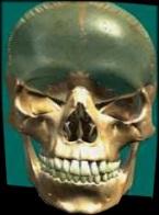
This translucent skull was mapped so as to view internal structures
in a fly-through for first year medical students.
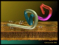
| The protein causing whooping cough was animated--- uncoiling,
opening calcium channels and going through the cell membrane to cause disease.
This was produced to support a feature NEW to academia at the time: animations
targeted at grant proposals in the basic sciences, the models of which were re-useable, modifications based on furthering research. This concept has caught
on in other areas as well, and digital media are now regularly submitted as appendices
to grants across the sciences. The domains in the protein were merely shown in symbolic colors to reduce computing time; current animators have the luxury of processing more elaborate protein envelopes, as computing has evolved over 1000x since that time.
|
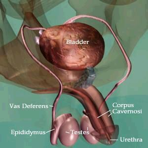
From an early QTVR of the Prostate and pelvis, the model is viewable from any attitude.
©1996 Urologic Multimedia, Inc.
CONTACT the
Artist
RiverScapes Gallery
All FineArt Bio
of the Artist
Medical
Illustration Commissions Medical
Illustration
All images ©1977-2011 CALuce, All rights reserved.